A research paper published in Science Advances has made new strides into the world of dynamic visualization of biological phenomena in cells. The study involved a new microscopy technique to label and observe live cells in order to record over time the interactions between DNA and proteins within the cells. An interdisciplinary team worked on a new and unique approach towards the visualization of the process within the cell that is used to make proteins from DNA.
DNA contains crucial information about the makeup of an organism. DNA contains the code that enables replication of necessary proteins in most life forms. It is the reason we are able to grow and maintain our bodies, through constant repair and regrowth. The methods of these processes are under careful watch by these scientists. Being able to properly visualize these processes could potentially help us come up with ways to prevent any errors in this process as such errors are what cause genetic diseases and some cancers.
A New Visualization Approach
Typically, most biological samples are observed as stills with stains to label specific portions of the sample under study. This new visualization approach however, pioneers the rarer methods of observation that involve taking dynamic visuals of samples in order to better visualize the processes occurring within these cells.
The research team led by Associate Professor Tim Stasevich consisted of scientists from the Departments of Biochemistry and Molecular Biology of Colorado State University. Statsevich said that this ability to capture a sequence of events as dynamic movies instead of static stills offers a new, powerful way to observe the impact chemical modifications have on proteins as they interact with DNA to accomplish the tasks essential for life.
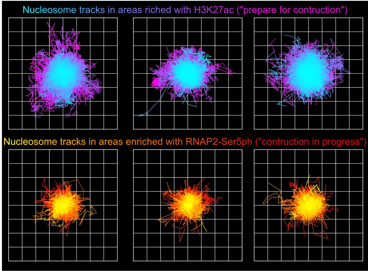
The team particularly focused their work in recording interactions around nucleosomes within cells. Nucleosomes are a mass formed by the wrapping of DNA around a histone protein core. The team wanted to record the interactions of Nucleosomes and the enzyme RNA polymerase II, which is the enzyme that catalyzes the process of mRNA formation from DNA during protein creation.

Image Credit: Science Advances (2023).
“We wanted to observe how genetic material responds when a cell activates transcription or marks areas for future transcription. This gives us much better information when we attempt to correct aberrant and disease-causing behavior,” Stasevich explained. Such work could one day help in controlling gene expression with applications in medicine, such as turning off genes that cause cancer, or preventing the manifestation of other such genetic disorders.
Results of the Challenging Research
The first author of the paper, Matthew Saxton remarked that it was rather difficult to pull off interdisciplinary research of this nature. It involved team efforts across the biochemistry biophysics and microscopy disciplines and a major amount of data analysis to pull off this challenge.
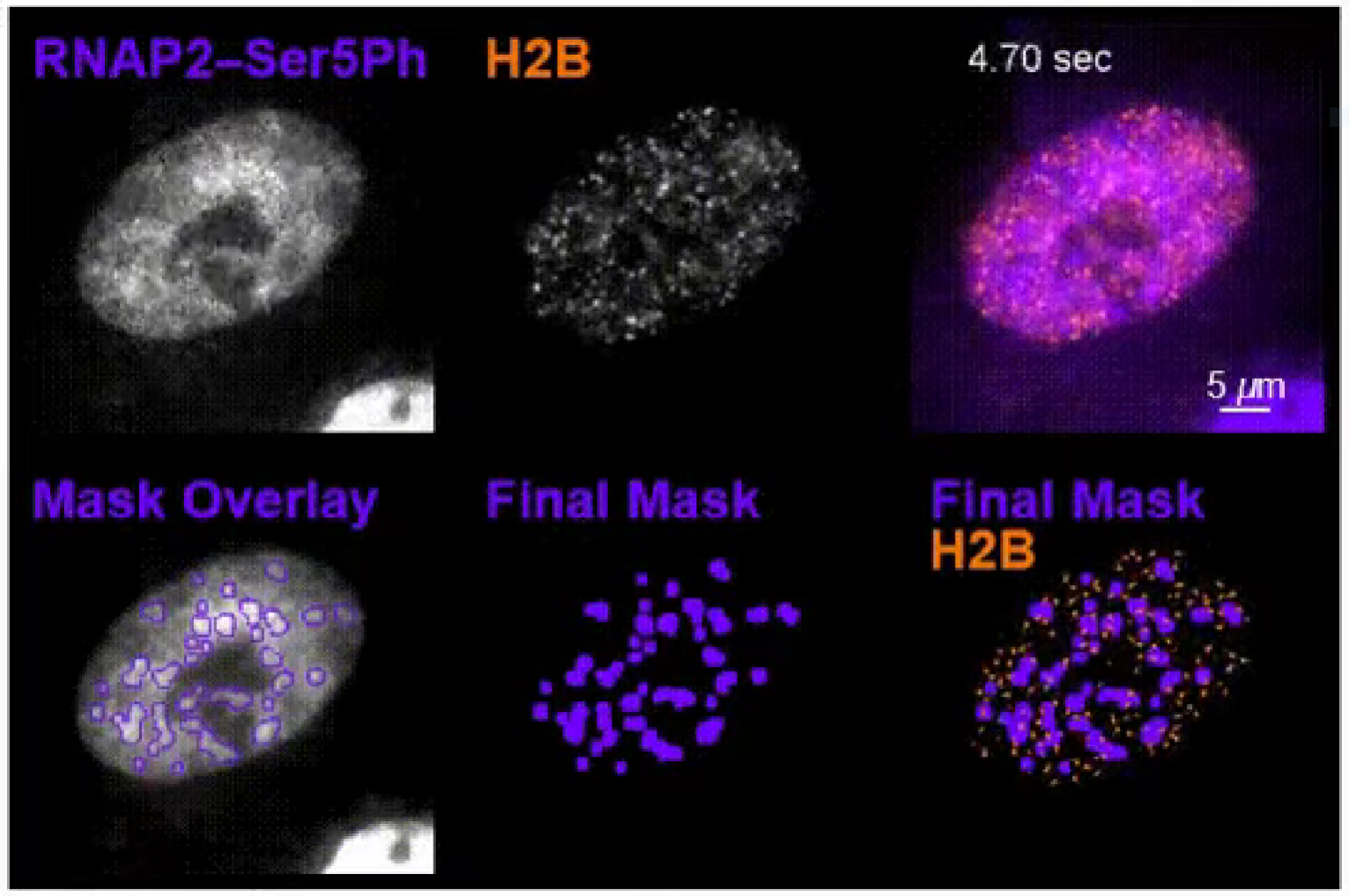
The cells had to be manipulated genetically with biochemistry and then imaged on a complex microscope using proper microscopy. And then the large volume of data that was output by the microscope had to be analyzed in order to obtain the dynamic render that they obtained at the end. The video generated from this visualization can be watched here.

Image Credit: Colorado State University.
He added that this research provides insights into how life functions, and can have a broad range of future applications. The research was aimed at observing how genetic material responds when a cell activates the mechanism to make protein from genetic material or marks areas for future proteins to be generated.
A look into these key biological processes can give a lot of information, potentially enabling correction of problematic and disease-causing behavior by cells during these processes.


